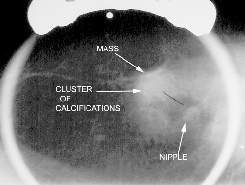
Calcification and mass abnormalities in breast mammogram scans
4.5 (657) In stock
Download scientific diagram | Calcification and mass abnormalities in breast mammogram scans. The calcification distribution depicts tiny flecks of calcium as small white regions on the left side, while the mass is shown as a smooth, well-defined border on the right side. from publication: Multi-Graph Convolutional Neural Network for Breast Cancer Multi-Task Classification | Mammography is a popular diagnostic imaging procedure for detecting breast cancer at an early stage. Various deep learning (DL) approaches to breast cancer detection incur high costs and are prone to classify incorrectly. Therefore, they are not sufficiently reliable to | Breast Cancer, Convolution and Classification | ResearchGate, the professional network for scientists.

Digital mammography dataset for breast cancer diagnosis research (DMID) with breast mass segmentation analysis

Breast mass, Radiology Reference Article

Atlas of breast cancer early detection

Invasive Breast Cancer Presenting as a Mass Replaced by Calcification on Mammography: A Report of Two Cases

Brendan JENNINGS, Head of Graduate Studies

Ductalca3.jpg

J. Imaging, Free Full-Text

a) The cropping breast profile image of mdb111 for left MLO

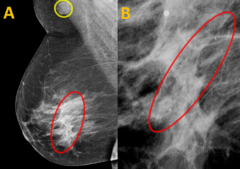
Cureus, Spontaneously Disappearing Calcifications in the Breast: A Rare Instance Where a Decrease in Size on Mammogram Is Not Good

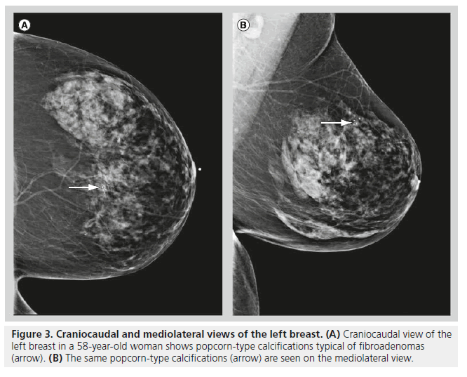
Mammography of breast calcifications

Shagufta HENNA, Lecturer

Early detection and classification of abnormality in prior mammograms using image-to-image translation and YOLO techniques - ScienceDirect
Dense Breast Tissue: What Does it Mean? - Mather Hospital
Less painful mammograms? New device may safely minimize the hurt
Small breast cancer, Radiology Case


